Digital X-Rays
Digital X-Rays
Digital X-rays are a form of imaging that shows what is happening beneath the surface of your smile: the roots, the jaw, and how everything is positioned. They help an orthodontist plan and monitor treatment with a clear view of the whole picture. This page explains how they work, how they fit into care at SS Orthodontics, and what a visit looks like.
What digital X-Rays is
An X-ray uses a very small, controlled amount of radiation to create an image of structures you can't see by looking in a mirror. Traditional X-rays captured that image on film, which then had to be developed in a darkroom before anyone could read it. Digital X-rays replace the film with an electronic sensor. The image appears on a computer screen within moments.
That change brings several practical improvements over film:
- Immediate images — the picture can be viewed right away, with no chemical developing step.
- Lower radiation — digital sensors generally need less radiation than film to produce a usable image.
- Adjustable views — brightness, contrast, and zoom can be changed on screen to study a specific area.
- Easy storage and sharing — images are kept as electronic files, which makes comparing them over time simple.
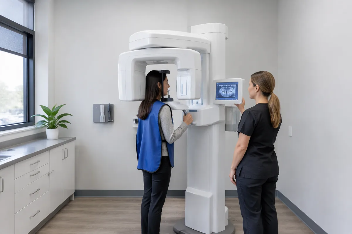
In orthodontics, the images most often discussed are a panoramic X-ray, which shows the whole upper and lower jaw in one wide view, and a side-profile head X-ray, which shows how the jaws and facial structures relate to each other.
How we use it at SS Orthodontics
Orthodontic planning depends on seeing more than what's visible in a smile. X-rays show the position of roots, whether anything is still developing or has yet to appear, and how the upper and lower jaws line up. That information helps an orthodontist decide what kind of treatment makes sense and how to approach it.
At our office, imaging is one part of the information-gathering that comes before a treatment plan. We also take digital scans, and after that our orthodontist creates a plan for your smile. The X-rays add to what the scans show by revealing what lies below the surface and within the jaw.
This kind of information is useful across many of the services we offer, including clear braces, metal braces, Invisalign clear aligners, early orthodontic treatment, and two-phase treatment. It is also relevant when jaw position is part of the picture, such as with surgical orthodontics, corrective jaw surgery, or TMJ-TMD concerns. Dr. Hoda Salim, our orthodontist, uses this kind of information when planning care.
What it means for you
For patients, the benefits of digital imaging are mostly about comfort, clarity, and confidence in the plan.
- Less radiation — digital sensors are known for requiring lower exposure than film, and the exposure from a typical orthodontic X-ray is small.
- Quicker answers — because images appear immediately, there is no waiting for film to develop, and a retake can be done on the spot if needed.
- Clearer conversations — seeing your own images on a screen makes it easier to understand what your orthodontist is explaining.
- A record over time — stored images can be compared with later ones to see how things are changing.
Dr. Salim's stated goal is for patients to feel comfortable, confident, and educated. Being able to look at your own images and ask questions about them fits that goal well.
What to expect during your visit
Before. There is usually little to prepare. You may be asked to remove items that could get in the way of the image, such as earrings, necklaces, or glasses. If you are or could be pregnant, tell our team beforehand so they can discuss it with you.
During. You will be positioned at the equipment and asked to stay still for a few moments. For some images you may bite gently on a small piece of plastic or rest your chin on a support. Capturing an image generally takes only moments. You may feel slight pressure from the positioning, but the X-ray itself cannot be felt.
After. The images appear on screen right away, and your orthodontist can review them. You can ask to see them and have them explained. There is no recovery time, and you can carry on with your day as usual.
Frequently asked questions
Are digital X-rays safe?
Digital X-rays use a very low amount of radiation, generally less than film-based X-rays. Protective measures such as a lead apron may be used. If you have concerns, especially about pregnancy, tell our team before any images are taken.
Do digital X-rays hurt?
No. The X-ray itself produces no sensation. Some people find that holding still or biting on a small positioner feels a little awkward, but it only lasts a short time.
How often will I need X-rays?
It depends on your treatment and your orthodontist's judgment. Images are commonly taken when a plan is being made and again at points when your progress needs to be checked. Ask our team what to expect for your situation.
Who are digital X-rays suitable for?
Imaging is used for children, teens, and adults alike, since orthodontic care is not limited to one age group. Children's images can show how growth and development are progressing. Adults' images help plan treatment around existing structures. Your orthodontist decides which images are right for you.
If you would like to know more about digital X-rays or how they fit into your care, get in touch with SS Orthodontics or book a visit, and our team will be glad to answer your questions.
This content is for general information only and isn't dental or medical advice. Talk with your dentist or orthodontist about your own situation. If you have a medical emergency, call 911.