Cephalometric X-Rays
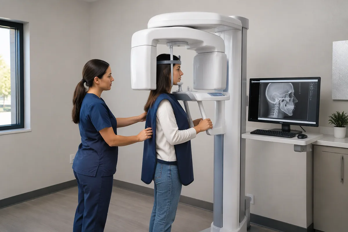
Cephalometric X-Rays
A cephalometric X-ray is a side-view image of the head that shows the jaws, the bite, and the profile of the face in a single picture. Orthodontists use it to understand how the structures beneath the smile fit together. This page explains what the image is, how it is used in planning care, and what a visit involves.
What cephalometric X-Rays is
"Cephalometric" simply means measuring the head. A cephalometric X-ray, often called a "ceph" film, is taken with the patient standing or seated in a fixed position, with the side of the face toward the imaging unit. The image captures the skull, the upper and lower jaws, the position of the teeth-bearing bone, and the soft tissue outline of the nose, lips, and chin.
What makes it useful is consistency. Because the head is positioned in a standard way, an orthodontist can mark reference points on the image and measure the angles and distances between them. These measurements describe how the upper and lower jaws relate to each other and to the rest of the face.
Before this kind of imaging, planning relied mostly on visual examination and hand-held measurements, which could not show what lay beneath the surface. A ceph image adds a clear view of the underlying bone structure. It also gives a baseline that can be compared with later images.
How we use it at SS Orthodontics
At SS Orthodontics, imaging is part of building a plan for your smile. Our introduction to the process says that we take digital scans, and then our orthodontist creates a plan. A cephalometric X-ray is a different kind of record from a digital scan, and the two answer different questions. Scans show the surface of the smile, while a ceph image shows the jaws and facial profile beneath it.
In general, a side-view image like this helps an orthodontist think through questions such as:
- Jaw relationship — whether the upper and lower jaws line up well front to back.
- Bite pattern — whether an overbite, underbite, or open bite comes from the position of the jaws, the position of the teeth-bearing bone, or both.
- Facial profile — how the lips and chin sit in relation to the structures behind them.
- Growth considerations — how a younger patient's jaws may be developing, which matters for early orthodontic treatment and two-phase treatment.
This kind of information is also relevant to several services we offer, including clear braces, metal braces, Invisalign clear aligners, and, when jaw position is a concern, corrective jaw surgery and surgical orthodontics. Whether a ceph image is needed for you is a decision for our orthodontist.
What it means for you
A ceph image gives you and your orthodontist something concrete to look at together. Seeing the jaws in profile can make it easier to understand why a bite looks the way it does, and why a certain approach is being recommended.
- Clearer planning — measurements from the image support decisions about how to move teeth and whether jaw position needs attention.
- Better communication — a visual reference makes explanations easier to follow, especially for parents of younger patients.
- A point of comparison — later images can be compared with earlier ones to see how treatment and growth are progressing.
- Few demands on you — the image itself requires no special effort. You hold still for a short moment.
What to expect during your visit
Before. There is usually little to prepare. Wear comfortable clothing, and remove earrings, necklaces, glasses, or hair accessories if asked, since metal objects can show up on the image. If you are or might be pregnant, tell our team beforehand.
During. You will be positioned with your head steady, often with small supports near the ears to keep the angle consistent. You will be asked to stay still, and sometimes to bite gently in your normal position or relax your lips. The exposure lasts only moments. Nothing touches the inside of your mouth, and most people describe it as simply standing still for a brief time.
After. The image is available to our orthodontist right away to review. There is nothing to recover from, and you can go on with your day. Your orthodontist can then use the image alongside other records to explain what they see and what it may mean for your plan.
Frequently asked questions
Is a cephalometric X-ray safe?
Dental and orthodontic X-rays use a small amount of radiation, and a single side-view image is a limited exposure. Protective measures such as shielding are commonly used in imaging. If you have concerns, ask our team how imaging is handled at SS Orthodontics.
Does it hurt?
No. The X-ray is taken from outside the mouth, and you only need to stay still for a moment. Some people find the head positioning a little unusual, but it should not cause discomfort.
How long does it take, and how often is it used?
The exposure itself takes only seconds, and positioning adds a short amount of time. How often a ceph image is taken depends on the individual case and the orthodontist's judgment. Some patients have one to help plan treatment, and others may have additional images to follow progress.
Who is it suitable for?
Cephalometric X-rays can be used for children, teens, and adults who are being evaluated for orthodontic care. They are especially informative when jaw position or growth is a question. Our orthodontist can tell you whether one makes sense for you or your child.
If you would like to know more about cephalometric X-Rays or how they fit into your care, get in touch with SS Orthodontics and our team will be glad to answer your questions.
This content is for general information only and isn't dental or medical advice. Talk with your dentist or orthodontist about your own situation. If you have a medical emergency, call 911.